Thursday, March 17, 2011

Bell's Palsy 101: An Introduction to the Seventh Cranial Nerve

There are 12 cranial nerves. They are so named because they come off of the brainstem before the brain & brainstem leave the skull to become the spinal cord. They control things such as sight and movement of your eyes, smell, feeling and movement of your face, etc. Number 7, the Seventh Cranial Nerve, is primarily responsible for the movement of your face. Your brain thinks about moving your face and then the seventh cranial nerve relays that message to the muscles. It does some other things as well as you'll see below, but movement of your face is the major function (and most noticeable to other people when it's not working).

Okay, there are facts about Bell's Palsy below, but I think we should start with a basic anatomy lesson first. In this first image, I can appreciate that it looks complicated, but the basics are straight forward if you just read this through. On the left (the purple spot) is the nucleus, the start, of the seventh cranial nerve, the nerve which is the source of all your trouble. I circled and numbered some of the nerve branches that lead to your symptoms:
1. This leads to the tiniest bone in the human body in the ear, which helps soften sound so they will be comfortable. When not working properly sounds are louder in the affected ear.
2. When not working properly/inflamed, it causes pain behind the affected ear
3. Involvement of this is what contributes to dryness of the eye/eye discomfort and leads to the biggest complication of Bell's palsy, corneal abrasion, and potential vision loss if lubricating eye drops and a patch are not used.
4.This is what causes an unusual taste (or lack thereof). This nerve helps supply taste to the front part of your tongue.
5. This helps with keeping your mouth moist.
6. Last but definitely not least, this is where the actual weakness come from. All these branches weave into the upper and lower face to provide strength. Some people report "sensation change" but they are actually experiencing an unusual sense of "fullness" in the mouth because of the weakness rather than true sensation change. Other people have true "tingling" as the length of the nerve is affected by the inflammation. Other people truly do have additional sensation change because an entirely different nerve (the Fifth Cranial Nerve) can sometimes be affects as a kind of collateral damage.

I went through all that so you could see that depending on which of these symptoms you have, you can see potentially what part of the nerve is affected in your personally.

The second picture below in color shows the same nerve with some of the branches taken away for simplification, overlying a face to help orient us. Please note that the picture is also reversed, with the start of the nerve on the right in the second picture. Also, they put in some nearby nerves: Six, Five, and Eight so you can see that they're close by.


What are the first two things I think of when I see someone with weakness one side of the face in the ER or in my office?
1. Stroke
2. Bell's Palsy

**** I care about whether you can lift your eyebrow****
Patients always look at me funny when I focus on this during my exam, but it's important. Lifting your eyebrow is controlled by both sides of the brain. So you have a stroke on one side of your brain that makes your face weak, the other side of your brain will take over to help lift your eyebrow at least. BUT if the seventh cranial nerve is damaged after it leaves the brain, neither side of your brain can get to the eyebrow to lift it. Therefore a gross rule is this: Able to lift eyebrow=possible stroke. Can't lift eyebrow=possible Bell's palsy.

If it's a stroke, I have to decide where it is since that changes treatment options and workup quite a bit.
If it's a pure Bell's palsy, I have to decide how bad it is, what treatment to do depending on timing, and prognosis for recovery. Growing evidence implicated Herpes simplex virus type 1 as the most likely culprit in this.

I say pure Bell's palsy because a palsy of the seventh cranial nerve can potentially (depending on the patient and history) be caused by quite a few things: A run-amok ear infection, trauma, Lyme disease, Ramsey Hunt Syndrome, neoplasm/tumor.
And if you have bilateral seventh nerve palsy or a return of your facial weakness in the future to the opposite side, or other nerves affects such as those nearby ones in the picture above, I have to also consider all the above but also such things as: bad luck and you just got a pure Bell's palsy on the opposite side, Guillain-Barre syndrome/Miller Fisher syndrome, multiple cranial neuropathies for other reasons, pseudotumor cerebri, brainstem encephalitis, meningeal carcinomatosis, syphilis, AIDS, TB meningitis, pontine hemorrhage, lupus, Sjogren's syndrome, amyloidosis, diabetes, eclampsia if your pregnant, flu vaccine, neurosarcoidosis....

Bell's palsy is almost always straight forward, but you can see it can be quite exotic too, which is why you shouldn't just ignore it.


Facts:
25 out of 100,000 people get Bell's palsy...but accounts for about 75% of all cases of one-sided facial weakness. So if you do get this weakness, probability is in your favor for it being this relatively benign process.
Most people have complete resolution within 3 to 6 months, although some people can have a subtle residual evidence on their driver's license photo when checked.
About 10% have recurrence.
Incidence is highest in those over age 70, and you have about the same chance of getting from age 30 through age 69. It's less common below the age of 29.
Most patients recover completely.
You have less of a chance of recovering completely if you are older, have hypertension, have impairment of taste, have pain other than behind the ear, and have complete facial weakness, have symptoms that progress more rapidly to fuller weakness over say less than 5 days than those that gradually get worse over more than 5 days.
The two most common symptoms (after slight residual weakness of course) include loss of vision if the production of tears is affected and the patient doesn't lubricate the eye appropriately, and something called "crocodile tears." Because there is sometimes damage to some of the nerves that go to the salivation gland, they can regrow incorrectly and connect to nerves responsible for tear production. The result is that when you have a salivary stimulus (you're hungry and a big tasty meal is placed before you), you may make tears. This saying comes from the fact that crocodiles are considered heartless predators and therefore "cry" while eating their tasty prey.

Treatment:

1.The best treatment for most people is tincture of time.
2. Studies are mixed about prednisone (taken at a decreasing dose by-mouth over 5 to 7 days) and antivirals such as acyclovir or valcyclovir. Taken within the first 24 to 48 hours of the start of the symptoms, most of us feel this is more likely to provide benefit in helping to reduce the severity of symptoms and possibly shorten the duration... although this is debated. It is agreed upon that taken after the first 24 to 48 hours, these medications are less likely to help.
3. Lubricating eye drops and an eye patch. Protecting the eye until it can close fully (at night as well) is essential in literally keeping your eye. Scratching the eye by rubbing against your pillowcase, your sleeve, etc. can quickly lead to infection and losing that eye.
4. Over the counter pain medications if you have pain in the distribution of the nerve (Aspirin, ibuprofen, Tylenol)
5. Moist heat applied to the face multiple times a day or before bed may help with the pain.
6. Physical therapy self-massage of the face and facial exercises may be helpful, and physical therapy is essential if you have additional damage to the nearby Eighth cranial nerve which can affect balance and walking.
7. B vitamins (B12 & B6) and zinc may help: They promote nerve health/growth.

Less likely to be helpful: Nerve decompressive surgery, biofeedback, acupuncture

There is so much more that could be said about Bell's Palsy. I hope this is a good start for you.

    What do you do as a Neurologist, exactly?

    Usually patients show up knowing exactly why they're here to see me, but sometimes patients show up and are not sure exactly what they are supposed to get out of this visit, what exactly I do as a Neurologist. It is not uncommon for someone to ask what type of patients I see. We as Neurologists see a wide spectrum of illnesses, anything that can directly, or often indirectly, affect the brain or spinal cord (central nervous system) or the nerves after they exit the spinal cord and weave their way into every nook and cranny of your body (peripheral nervous system). You can imagine that this would encompass a wide variety of illnesses such as the headline-dominating illnesses like Strokes and Alzheimer's disease, Parkinson's and Epilepsy, Tumors and Multiple Sclerosis among many many others... not to mention all the exotic and mysterious multi-syllabic-named illnesses out there, and the very common complaints such as carpel tunnel syndrome, headaches, neuropathy in the feet.

    From day-to-day, when I'm not seeing patients in the hospital, I'll see an average of 5 to 8 new outpatients a day, and the rest are follow up on our prior visits or studies such as MRIs/lumbar punctures/urine studies/blood work, sleep studies or electroencephalograms (EEGs) to explain the results which can often be very tedious because of the diagnoses we are considering. I spend an average of 45 minutes with each new patient currently (although current healthcare cuts may make us change to 30 minute visits in the future). So in a 5 day week, I currently see an average of 25 to 40 new patients and a whole bunch of follow ups.

    I wanted to give you a touch of insight into what people complain of, so I took word-for-word, the chief complaints patients write on their "REASON FOR THIS VISIT" section of their form and list for you a whole week's worth of new patient subjective chief complaints. This is what I see as their complaint before I walk in to see what I can do to help address it:

    (Age)-----(Complaint)
    43-----I feel off
    76-----Trouble speaking
    80-----Memory decline
    36-----Neurocysticercosis and sleepy
    20-----Narcolepsy
    50-----Excessive daytime fatigue and sleepiness
    23-----Night fits
    63-----Neck pain and hands numb
    45-----I snore like the banshee
    40-----Post traumatic hypersomnolence
    29-----Cognitive problems, fatigue, joint pain, muscle pain, insomnia, anxiety
    85-----Imbalance
    52-----Increasing weakness; cerebral palsy
    33-----Multiple sclerosis?
    60-----Difficulty sleeping
    75-----Weakness in arms and legs
    65-----Recent "mini stroke"
    45-----Sudden loss of consciousness
    60-----PTSD at night? Kicking a lot.
    51-----My vision is "shaky"
    45-----Worms feeling in my legs
    37-----Head injury-weakness
    37-----Discomfort in neck, left arm, left leg, getting worse
    76-----right leg weakness
    66-----TIA
    39-----Bell's palsy?
    35-----Burning in my toes
    58-----Vertigo
    56-----Severe restless legs- meds don't work on me
    74-----Gait disturbance
    71-----Memory poor
    35-----Epilepsy
    43-----Off balance, speech pattern
    67-----Sensation changes in arms and legs
    53-----Continued 1 month right-sided headache
    74-----Passing out but aware of surroundings
    26-----Severe headache, pregnant

    Sunday, March 13, 2011

    Tinnitus 101: A Basic Introduction

    When most people think of tinnitus, they think of ringing in the ears. But actually, the sounds that make up tinnitus are broken down into the following:
    1. Ringing (37.5%)
    2. Buzzing (11.2%)
    3. Cricket-like (8.5%)
    4. Hissing (7.8%)
    5. Whistling (6.6%)
    6. Humming (5.3%)

    In some people it is loud and in others soft. An average of 34% say it is 8 or more on a 10-point loudness scale. It is more common as people age and is more common in men than in women. In fact, almost 12% of men age 65-74 are affected.
    The differential for what causes tinnitus is actually large, but most people (and many clinicians) fail to recognize that it is important to differentiate subjective tinnitus (what you the patient appreciates) versus objective tinnitus (something that can actually be heard by something else like another person or more likely sensitive otoacoustic equipment). This is because the actual diagnoses that fit into subjective versus objective tinnitus differ.

    Examples:
    Subjective tinnitus heard only by the patient. Causes:

     Neurologic: Head injury, whiplash, multiple sclerosis, acoustic neuroma tumor, stroke, other tumors where the cerebellum meets the pons (brainstem)
    Drugs such as aspirin, NSAIDs like Aleve and Motrin etc., antibiotics (aminoglycosides), loop diuretics for hypertension, chemotherapy
    Infectious: Ear injections, prior Lyme disease, meningitis, syphilis, etc.
    Otologic: Long-term noise exposure, presbycusis (natural aging of the ear), cerumen impaction (ear wax), Meniere's disease, etc.

    By the way, digression here: I should mention here that if I diagnose tinnitus in an individual older than 75, I consider first a tumor, then stroke (both of which can usually be ruled out with an MRI at once) and then medication changes or ear infections (viral or bacterial) then most likely age-related changes of the ear itself which just happens sometimes. Sometimes, in the elderly, there can be narrowing in the vessels leading to the head, or in the head itself, which can cause a whooshing sound due to turbulent flow of the blood.

    Objective tinnitus heard by more than just the patient. Causes:
    Spontaneous: Spontaneous otoacoustic emissions (your ear just makes music for the world by vibration of the outer hair cells of the cochlea-- this is actually very common and most people have randomly experienced this from time-to-time)
    Muscular: Palatal myoclonus (usually due to brainstem damage causing this secondarily), spasms of the stapedius or tensor tympani muscle (the tiniest muscles in the ear than normally protect the ear from noises too loud), etc.
    Pulsatile (comes and goes rhythmically): Carotid stenosis (narrowing of your carotid arteries), Vessel tumors or abnormal formations of the blood vessels either locally in the ear/brain or in the vessels that lead to the ear/nearby brain, valve abnormalities in the heart (such as the aortic valve which is the last valve before blood leaves the heart.


    I like this drawing below, because it shows some of the pathologies mentioned above that can cause tinnitus and their locations (compared to the brain and nose's location). The first drawing is a small one showing you the ear and some the basic anatomy so you can compare it.


    Normally, tinnitus has a straight forward cause and easy to diagnose a cause through our discussion and exam, even if I order some additional test to rule out one of the more serious possible causes (since we don't want to miss these of course).
    When people do have tinnitus, 22% experience it in both ears equally, 34% only in one ear, and the remainder in both ears but with one ear being louder. So you can see that only a third of people have it in only one ear. That does not mean there is a tumor in that one ear or something happening only in that ear; we just have to consider that possibility.
    Questions to help us decide what's going on:
    Is it constant or episodic?
    Both ears or just one?
    Sudden onset or gradual?
    How long has it been happening?
    How loud is it?
    How high is the pitch?
    Are there other conditions?
    Anything make it more likely such as background noise, alcohol, stress, sleepiness?
    Do you have a history of noise exposure, ear infections, ear surgeries, ear deafness in the past, head injuries, recent medication changes, other things that happen at the same time?
    Pain in that ear(s)?
    Does it affect your daily living or ability to function?

    The most common things I do to delineate the cause besides a history and head/neck exam (if needed):
    MRI brain +/- MRA of the vessels, echocardiogram, audiologic evaluation, ENT referral

    Treatment (assuming a benign cause): That's what everyone really cares about.
    Unfortunately, although there are many medications that can cause tinnitus there are not any FDA-approved medications to treat tinnitus directly although some are used indirectly such as:
    Tricyclic antidepressants ( amitriptyline and nortriptyline) These have been used with some success. But these medications are generally used for only severe tinnitus, as they can cause side effects like dry mouth, blurred vision, constipation, heart problems, and confusion in the elderly.
    Alprazolam (Xanax) may help take the edge off the symptoms, but side effects can include drowsiness and nausea, and they can become habit-forming, or increase fall risks in the elderly.

    Other treatments:
    Noise suppression to "out compete" the tinnitus, such as white noise machines (falling rain, city traffic, ocean waves).Hearing aids if it present in people with associated hearing loss. Masking aids worn in the ear which essentially are white-noise-machine-meets hearing-aid and produce a low-level white noise in the ear during the day.Tinnitus retraining which is a wearable device that plays tonal music that over time helps one not to focus on the tinnitus.

    Alternative medications:
    Despite numerous trials utilizing ginkgo biloba, no firm conclusion regarding efficacy is yet reached. It is relatively benign at usual treatment doses and a dose 120-160mg should be tried if desired.

    Other lifestyle strategies:
    Decrease alcohol (it increases inner ear blood flow and can make tinnitus worse).
    Decrease stress and get a normal amount of quality sleep.
    Treat any underlying depression.
    Add gentle background noise to your day (fan, soft music, low volume radio static).
    Stop smoking (nicotine is an irritant).
    Stop listening to loud music/exposing yourself to other loud sounds, or wear ear plugs if you cannot avoid it.

    Thursday, March 10, 2011

    What's Happening in my PSG/Sleep Study?


    A patient came in and brought a CD with a prior sleep study and based on their symptoms I took a look at it later and I thought this might be a good chance to take a screen shot and then show it here so you can see what an average screen looks like so you can see what I review after you get your overnight study.
    This screen shot represents 30 seconds, and the average person sleeps about 6.5 hours during their sleep study, so I review on average 780 of these screen shots each time I review a study. It goes quicker than you think, mainly because I've looked at so many now.
    ---First, at the bottom, you can see a time-of-night bar which shows you where you are in the night. You can see on here that we are early in the night (23:10:38 to be exact if you look closely there or in the upper left corner).
    ---Next, you can see see the "SaO2" which shows blood oxygenation. This person is averaging 94 and 95%.
    ---Above that you can see "Abdomen" and "Thorax"  which are from the bands around your stomach and chest. They show movements which help characterize your breathing since there are different type of abnormal breathing patterns (i.e. obstructive apneas, central apneas, hypopneas, mixed apneas, Cheyne-Stokes... etc.)
    ---The "Flow" above that also helps us via assessment of airflow in your upper airway.
    ---Next are the "RAT" and "LAT" which assess your leg movements at night... periodic limb movements (PLMS) which can be associated with restless leg syndrome (RLS) or independent, but other abnormal movements of the limbs can be evaluated by these leads as well.
    ---The "ECG" checks your heart with one lead, and checks basic rhythm and gives a gross view of any premature beats/missed beats/how they may change during the night. Some people have sudden changes associated with their respiratory events.
    --- "ROC" and "LOC" tell us what your eyes are doing and helps immensely with staging of your sleep such as slow wave sleep versus REM (dreaming) sleep.
    --- "Snore" is self-explanatory. It's a little mic that picks it up.
    --- The "Chin" lead tells us if you're having muscle movements in your chin and helps us stage sleep and look at other things like bruxism (clenching/grinding of your teeth at night)
    --- The top 6 lines: "Cz-T4" "T3-Cz" "Fp1-O2" "O1-A2" "C3-A2" are the EEG electrodes placed on your head during the study. You don't have to have all the ones we have here, but because I'm a Neurologist, I pay closer attention to EEGs and often get referrals for things that happen at night concerning for possible nocturnal seizures versus other things which a few more EEG leads help you differentiate. This is the most interesting part of this particular Polysomnogram (sleep study) actually: You can see about 2/3rds of the way across that these leads show some large amplitude waves. If you look at them closely, you can see that there is a "wave" and a jagged "spike" component to each of them (maybe 8 or 9 of them total). We call these... "spike and wave" complexes. These are actually consistent with a generalized seizure this patient had during this study. Now I would need a full EEG with many more leads to characterize better... but this person didn't have breathing problems, but instead was feeling awful during the day because they kept having small seizures throughout their night.
    I hope you found this interesting. I sure do.

    Monday, March 7, 2011

    Ridiculous... Treatment for Snoring and other Sleep Problems

    I am always seeing ads for different sleep aids, and patients are always bringing them in to ask me if it's worth their time or money. Some of them are downright hilarious:

    I call this "The Bull."
    I think it's self-mocking enough... and completely ineffective.
    (It's also 10 cents worth of plastic)


    Okay, now these are meant to be cute pillows I assume, but number 2 looks more like a pair of underwear. And is it really going to pull your jaw forward? How? Even custom made dental appliances have a hard time of doing this.


    This should be called the "Cranky Spouse."
    So... you snore, which wakes you up and disrupts your sleep. So we're going to solve this by waking you up with shocks.
    It reminds me suspiciously of the shocking dog collar "invisible fence". In this case, it's the invisible boundry of snoring tolerated by your spouse.


    It looks like this man is blowing a bubble with his gum.
    What I find most amusing about this is that we all can see that it looks completely ridiculous, but they try to smooth that over with an attractive wife giving us the thumbs up. 
    To be honest, I actually do not know if this would work or not. Maybe for someone with a big tongue who has apnea only to a mild degree in the supine position.


    I actually like the one on the right.
    The one on the left looks like the set up at the massage therapist's office.
    They aren't trying to make any apnea claims here it seems, but I do think the model on the left was probably disappointed when she showed up for the shoot.


    Again, no claims about sleep, but this does kind of look like a pillow from Space Odyssey 2001.
    Or as though someone is storing their head like wine. 


    This one frustrates me a little.
    Not because as a person who reads EEGs, this is making the art of EEG interpretation a seemingly juvenile endeavor.
    Not because I know it can't properly identify "sleep stealers."
    Not because your "ZQ" is ridiculous, especially because I know their score is useless.
    But because they try to charge you $249 dollars, less than you have to pay (assuming you have insurance) for a complete overnight sleep study in a lab with all the bells and whistles, a sleep technologist there with you all night, and the physician sleep specialist looking over your study to personally interpret your findings.


    This CPAP mask actually works for some people. For people who truly feel claustrophobic while wearing their mask despite many different harder ones tried, or hate the actual hardness itself, this mask can be useful in keeping patients compliant and therefore helped by the use of CPAP.

    But look at it. Objectively, it's ridiculous.
    And leopard print?
    

    Seriously?.. Leopard print?

    Sleep Disorders are Simple and They're Not

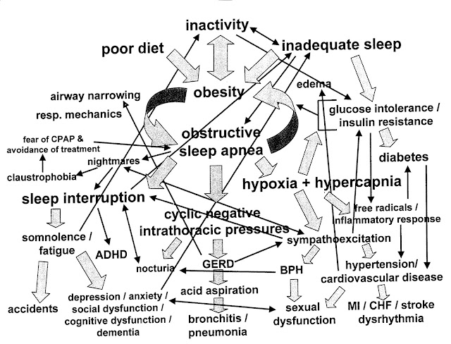
    This diagram was given to me by a new sleep patient once. It was given to them by their prior sleep specialist at their new visit with them. Many years later, they were still suffering from poor sleep and excessive daytime sleepiness from what turned out to be a relatively straight forward cause. We were able to address it without even needing a new sleep study, just a long conversation discussing their sleep issues. Anyway, I asked them if I could make a copy because it made me laugh. How intimidating is this chart?! Why would I show someone this? It looks like the slide from some scientific meeting.


    I decided that if you could understand that diagram, you could probably work this and then procede to fly off in it after I showed you the above diagram:


    The reality is that yes, Sleep Medicine can be very complex. It is at the intersection of many medical disciplines: Neurology, Pulmonology, Cardiology, Otolaryngology, Psychiatry, and the implications of different sleep disorders (More than 85 different sleep disorders last count) cross-affects many different medical specialties.
    But, many of these sleep disorders can be evaluated and diagnosed without requiring an overnight sleep study.
    If you do need a sleep study, we essentially look at the following basic parameters:


    The summary is actually pretty straight forward at this level. From top to bottom we see:
    -Sleep stages (notice the stage REM dreaming in black)
    -Body position (This person started on their right, then went to their back, then left... never on their stomach)
    -Oxygenation (We want it to stay close to 100%. This person dropped severely)
    -Respiratory events (There are many types of respiratory events. This person had mainly two types but many of them)
    -CPAP (You can see it starts in the latter part of the night, and we increase the pressure to try and find one that stops or markedly improves the number of respiratory events. This person had a trend of improvement but may need more work)
    -Arousals (This is arousals from all different types of things: breathing issues, leg movements, pain, bed partner snoring... although we have to figure some of this out more by discussion than this estimate alone. This at least tells us a basic idea of what all woke you up this particular night)
    -Periodic Limb (leg) Movements during your sleep (not a problem for this individual)

    We can check many other things as well, such as carbon monoxide levels, or we can add a full EEG to your head to look not only for sleep stages with our basic EEG, but for actual nocturnal seizures. We also do other tests for things such as Narcolepsy or for more complex sleep apnea or assessment of daytime wakefulness.

    But in your average initial diagnostic polysomnogram (PSG), the fancy word for sleep study, these are the basic things we look at and utilize to help determine how an average night of sleep unfolds for you.

    So I hope when you go to discuss these things with your sleep specialist physician, you can just relax and tell him or her what is going on, and let them figure out where to fly from there.